Description
The Coomassie brilliant blue staining method is often used in the laboratory to stain the isolated proteins on the gel. It not only overcomes the limitation of the low sensitivity of amino black staining, but also is easier to operate than silver staining. Coomassie brilliant blue is widely available in G250 and R250. Coomassie brilliant blue G250 is often used in the determination of protein content because of its rapid binding reaction with protein. Coomassie brilliant blue R250 reacts slowly with proteins, but can be eluted off, so it can be used to stain electrophoresis bands.
This product is designed based on the Coomassie brilliant blue staining method for protein staining. Compared with traditional stain solution, this product is a new type of PAGE dyeing reagent, which does not contain toxic or irritating substances such as methanol and acetic acid. Its unique formula enables the function of surfactant stripping, protein fixation, protein staining, shielding non-protein area coloring at the same time. 8-10 minutes to finish PAGE protein staining without decolorization. The resolution of dyeing is equivalent to that of traditional staining methods. The stained protein bands or protein spots can be used for high efficiency electroelution and are suitable for protein spectrum analysis.
Features
- Does not contain toxic or irritating substances such as methanol and acetic acid.
- 8-10 minutes to finish PAGE protein staining without decolorization.
- Rinsed several times with clean water on a rotating shaker, the residual weak background can be completely removed.
Applications
- Protein staining
Specifications
| Detection Location | In-Blot Detection, In-Gel Detection |
| Label or Dye | Coomassie |
| Product Type Specs | Protein Gel Stain Reagent |
| Target Molecule | Protein |
Components
| Components No. | Name | 20309ES03 |
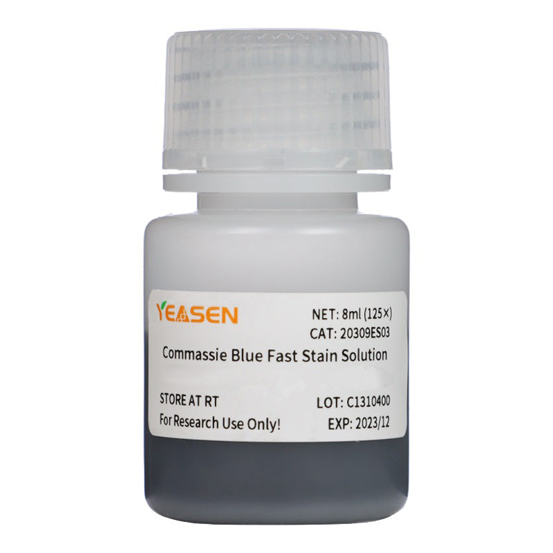
| 20309 | Commassie Blue Fast Stain Solution (8 mins) | 8 mL(125×) |
Storage
The product should be stored at room temperature for one year.
Figures
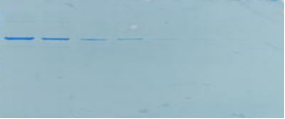
- Demonstration of commassie blue fast stain solution

Figure 1. The staining effect. The concentration gradient of BSA sample was 1000ng, 500ng, 125ng, 80ng, 20ng, 10ng from left to right.
Payment & Security
Your payment information is processed securely. We do not store credit card details nor have access to your credit card information.
Inquiry
You may also like
FAQ
The product is for research purposes only and is not intended for therapeutic or diagnostic use in humans or animals. Products and content are protected by patents, trademarks, and copyrights owned by Yeasen Biotechnology. Trademark symbols indicate the country of origin, not necessarily registration in all regions.
Certain applications may require additional third-party intellectual property rights.
Yeasen is dedicated to ethical science, believing our research should address critical questions while ensuring safety and ethical standards.


