Description
UltraNuclease, also known as a non-restrictive endonuclease, broad-spectrum nuclease, is a non-specific endonuclease derived from Serratia Marcescen, which hydrolyzes internal phosphodiester bonds between any nucleotides in nucleic acids to produce 5'-monophosphate oligonucleotides of 2-5 bases in length. It can degrade DNA and RNA in various forms (double-stranded, single-stranded, linear, circular, native or denatured) under a very broad range of conditions (6 M Urea, 0.1 M Guanidine HCl, 0.4% Triton X-100, 0.1% SDS, 1 mM EDTA, 1 mM PMSF) and is widely used to remove nucleic acids from biological products. UltraNuclease can also be removed by corresponding methods subsequently.
This kit uses the principle of double-antibody sandwich enzyme-linked immunosorbent assay (sandwich ELISA) to detect the residues of denatured and non-denatured UltraNuclease. First, coat the microplate with an anti-UltraNuclease rabbit polyclonal antibody to form a solid-phase antibody. Second, add UltraNuclease standard and test sample to the solid-phase antibody microplate, then add biotin labeled Anti-UltraNuclease polyclonal antibody, and finally, add horseradish peroxidase-labeled streptavidin (SA-HRP) to form an antibody + antigen + antibody-Biotin + SA-HRP complex. Subsequently, TMB substrate was added to the complex to observe color reaction after washing the complex. TMB is converted into blue under the catalysis of HRP enzyme and finally converted into yellow in the presence of acid, and the shade of color is positively correlated with the amount of UltraNuclease in the sample.
The detection quantification range of this kit is 0.047-3 ng/mL; the lower detection limit is 23.5 pg/mL.
Feature
- Highly sensitive: Detecting as little as 23 pg/ml of UltraNuclease residuals in in-process and final biological samples.
- Ensures accuracy: The UltraNuclease residuals can be accurately detected with a recovery of 80%-110%.
- Specificity: Cooperate use with YEASEN UltraNuclease and can accurately detect the residuals.
- Stable: The difference between lot-to-lot is low, the kit property is not impacted under 37℃ for 6 days.
- Easy signal collection: Using Biotin-Streptavidin system, the signal can be stably enlarged.
- Complete kit: All key components are provided in the kit, it’s Cost-effective for use.
Application
- Residual UltraNuclease test in biological products
Specification
| Sensitivity | 23 pg/mL (range 0.047~3 ng/mL) |
| Assay Time | <4 hours |
| Assay Principle | Two-site immunoenzymetric assay |
| Signal Amplification | Biotin-Streptavidin system |
| Detection Wavelength | 450nm |
Components
| Components No. | Name | 36701ES59 (96 T) |
| 36701-A | ELISA Microplates | 1 Plate |
| 36701-B | Detection Antibody: Biotin-conjugated Rabbit Anti-UltraNuclease Antibodies | 35 μL (0.2 mg/mL) |
| 36701-C | Standard: UltraNuclease | 1 vial (500 ng/mL) |
| 36701-D | HRP-conjugated Streptavidin | 10 μL |
| 36701-E | Dilution Buffer 1 | 45 mL |
| 36701-F | 20×Wash Buffer | 50 mL |
| 36701-G | Dilution Buffer 2 | 30 mL |
| 36701-H | TMB | 15 mL |
| 36701-I | Stop Solution | 10 mL |
| 36701-J | Sealing Film | 5 pieces |
Shipping and Storage
The UltraNuclease ELISA kit is shipped with an ice pack and can be stored at 2°C ~8°C. Unopened product is valid for one year. Once the reagent is opened, it is valid for half a year. Never freeze this product.
Storage of the prepared reagents:
1. The diluted washing buffer and dilution buffer can be stored at 2°C ~8°C for 1 week
2. Standard 36701-C is liquid and stored at 2°C ~8°C for 1 year
3. Prepared detection antibody and stop buffer can be stored at 2°C ~8°C for 1 month
Figures
- Specificity Demonstration

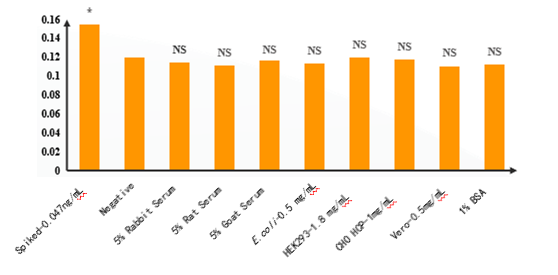
Figures 1. The specificity of UltraNuclease ELISA kit was shown in the figure above.
8 mimic samples were estimated for the non-specific reaction with the antibodies in the kit. There was no significant difference between the 8 samples and the negative sample. (*: Significant difference from Negative sample;NS: No significant difference from Negative sample)
- Precision Display
A)
| Concentration (ng/mL) | 6 duplicated data in the same plate | CV | |||||
| 3 | 2.412; | 2.390 | 2.323 | 2.367 | 2.476 | 2.291 | 2.53% |
| 1.5 | 1.780 | 1.883 | 1.884 | 1.908 | 1.876 | 1.883 | 2.19% |
| 0.75 | 1.123 | 1.215 | 1.166 | 1.18 | 1.140 | 1.109 | 3.12% |
| 0.375 | 0.691 | 0.706 | 0.708 | 0.722 | 0.722 | 0.679 | 2.21% |
| 0.1875 | 0.449 | 0.462 | 0.453 | 0.460 | 0.447 | 0.433 | 2.13% |
| 0.094 | 0.306 | 0.308 | 0.304 | 0.311 | 0.295 | 0.302 | 1.65% |
| 0.047 | 0.242 | 0.241 | 0.240 | 0.241 | 0.256 | 0.240 | 2.37% |
| 0 | 0.183 | 0.192 | 0.189 | 0.187 | 0.189 | 0.189 | 1.43% |
B)
| Intermediate Precision | |||
| Conc. | 1.5 ng/mL | 0.375 ng/mL | 0.094 ng/mL |
| Analyst 1 | 1.388 | 0.382 | 0.079 |
| Analyst 2 | 1.458 | 0.363 | 0.068 |
| Analyst 3 | 1.318 | 0.37 | 0.063 |
| Average | 1.388 | 0.372 | 0.07 |
| SD | 0.070 | 0.010 | 0.008 |
| CV | 5.04% | 2.59% | 11.69% |
Figures 2. The precision of UCF.ME™ UltraNuclease ELISA was assayed as the above scheme.
6 duplicated were tested at each concentration on the standard curve, and all the CV% were lower than 5% (Figure 2A). 3 analysts conduct the UCF.ME™ UltraNuclease ELISA experiment and the CV% were lower than 15% (Figure 2B).
- Accuracy Validation
A)

B)
| Concentration | OD450 | Result | ||||
| ng/mL | Repeat 1 | Repeat 2 | Repeat 3 | Average | Conc. | Recovery% |
| 3 | 2.271 | 2.222 | 2.287 | 2.260 | 2.950 | 98% |
| 1.5 | 1.495 | 1.488 | 1.514 | 1.499 | 1.540 | 103% |
| 0.750 | 0.868 | 0.856 | 0.890 | 0.871 | 0.735 | 98% |
| 0.375 | 0.537 | 0.525 | 0.522 | 0.528 | 0.358 | 95% |
| 0.188 | 0.373 | 0.369 | 0.389 | 0.377 | 0.201 | 107% |
| 0.093 | 0.261 | 0.255 | 0.258 | 0.258 | 0.082 | 88% |
| 0 | 0.184 | 0.195 | 0.187 | 0.189 | 0 | 0 |
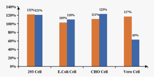
Figures 3. The accuracy of UCF.ME™ UltraNuclease ELISA was assayed as the following scheme.
UltraNuclease was spiked in 5 μg/mL cell lysis solutions (HEK293, E.coli, CHO, and Vero) independently with a final spiked conc. of 1.5 ng/mL. The accuracy in different cell samples showed excellent than other vendor(Figure 3A). UltraNuclease was spiked in an actual sample (provided by the client) and is serial 2 folds diluted from 3ng/mL to 0.093ng/mL. The spike recovery was all between 80%~110% (Figure 3B).
Payment & Security
Your payment information is processed securely. We do not store credit card details nor have access to your credit card information.
Inquiry
You may also like
FAQ
The product is for research purposes only and is not intended for therapeutic or diagnostic use in humans or animals. Products and content are protected by patents, trademarks, and copyrights owned by Yeasen Biotechnology. Trademark symbols indicate the country of origin, not necessarily registration in all regions.
Certain applications may require additional third-party intellectual property rights.
Yeasen is dedicated to ethical science, believing our research should address critical questions while ensuring safety and ethical standards.


