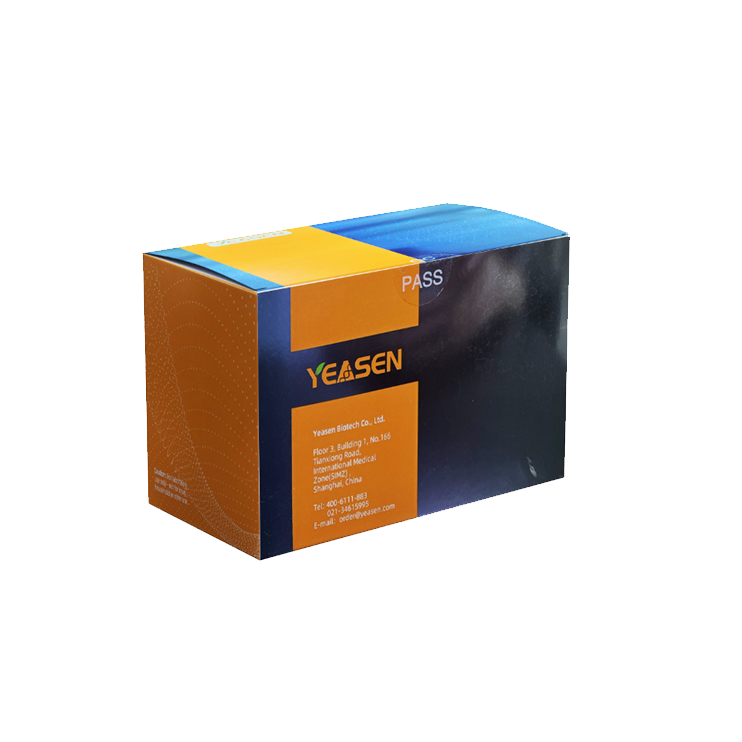
Description
Annexin V-EGFP/PI cell apoptosis detection kit uses EGFP-labeled Annexin V as a probe to detect the occurrence of early cell apoptosis.
The detection principle is as follows: in normal living cells, phosphotidylserine (PS) is located on the inner side of the cell membrane, but in early apoptotic cells, PS flips from the inner side of the cell membrane to the surface of the cell membrane and is exposed to the extracellular environment. Annexin-V is a Ca2+ -dependent phospholipid-binding protein with a molecular weight of 35 to 36 kDa that binds to PS with high affinity. The phosphatidyl serine can bind to the membrane of early apoptotic cells through the exposed phosphatidyl serine on the outside of the cell.
In addition, Propidium Iodide (PI) is provided to distinguish surviving early cells from necrotic or late apoptotic cells. PI is a nucleic acid dye, which can not pass through the intact cell membrane of normal cells or early apoptotic cells but can pass through the cell membrane of late apoptotic and necrotic cells and make the nucleus red. Thus, when Annexin V was combined with PI, PI was excluded from living cells (Annexin V-/PI-) and early apoptotic cells (Annexin V+/PI-). The late apoptotic and necrotic cells were double positive for both EGFP and PI (Annexin V+/PI+).
This kit is suitable for flow cytometry and fluorescence microscopy.
Product Components
|
Component |
|
40303ES20(20 T) |
40303ES50(50 T) |
40303ES60(100 T) |
|
40303-A |
Annexin V-EGFP |
0.1 mL |
0.25 mL |
0.5 mL |
|
40303-B |
PI Staining Solution |
0.2 mL |
0.5 mL |
1 mL |
|
40303-C |
1×Binding Buffer |
10 mL |
25 mL |
50 mL |
Shipping and Storage
The components are shipped with an ice pack and can be stored at -20°C keep out of light, and avoid repeated freezing and thawing for 1 year.
[Notes]: If it needs to be used repeatedly in a short period, it can be stored at 4 ℃ and protected from light for half a year.
Cautions
1) Since apoptosis is a rapid process, it is recommended that samples be analyzed within 1 hour after staining.
2) For adherent cells, digestion is a critical step. Do not use EDTA in the digestive solution as EDTA can affect the binding of Annexin V to PS.
3) Annexin V-FITC, but not Annexin V-EGFP, should be used to fix cells, such as to detect apoptosis and cell cycle at the same time, because EGFP would be denatured and lose its ability to excite fluorescence during fixation. Cells were incubated with Annexin V-FITC before fixation and unbound Annexin V-FITC was washed away with Binding Buffer. Because increased cell permeability during fixation creates cell debris that can bind to Annexin V and interfere with the results.
4) If the sample is from blood, be sure to remove platelets from the blood. Because platelets contain PS, which can bind to Annexin V, interfering with the results. Platelets can be washed away by using a buffering agent containing EDTA and centrifuging at 200 g.
5) Please centrifuge the reagent briefly before opening the cover, and throw the liquid on the inner wall of the cover to the bottom of the tube to avoid liquid sprinkling when opening the cover.
6) Annexin V-EGFP and PI are photosensitive substances, so please avoid light during operation.
7) For research use only!
Instructions
Experiment design
Blank tube: Negative control cells without Annexin V-EGFP and PI Staining Solution were used for voltage regulation.
Single stained tubes: Positive control cells, supplemented only with Annexin V-EGFP, were used to modulate compensation.
Detecting tube: The cells were treated with Annexin V-EGFP and PI Staining Solution. The experimental data were obtained by adjusting the parameters of the blank tube and single dye tube.
1.1 Sample dyeing
1) suspension cell: Cells were collected by centrifugation at 300 g for 5 min at 4 ° C.
adherent cell: After digestion with trypsin without EDTA, cells were harvested by centrifugation at 300 g for 5 min at 4 ° C. Trypsin digestion time should not be too long to prevent false positives.
2) The cells were washed with precooled PBS twice, each time at 300 g, and centrifuged at 4 ℃ for 5 min.
3) The cells were resuspended with 1×Binding Buffer and the concentration was adjusted to 1~5 ×10⁶/mL.
4) 100 μL of cell suspension was added into 5 mL flow cytometry tubes, 5 μl Annexin V-EGFP was added, and the cells were mixed and incubated for 5 min at room temperature under the light.
5) Adding 10 μl PI Solution and 400 μl PBS, the Staining was performed immediately.
[Notes]: When using flow cytometry to detect apoptosis, PI is greatly affected by time, and too long time will lead to an increase in PI staining, so flow cytometry should be completed within 1 h.
1.2 Flow cytometry analysis
The maximum excitation wavelength of EGFP was 488 nm, and the maximum emission wavelength was 507 nm; The maximum excitation wavelength of the PI-DNA complex was 535 nm, and the maximum emission wavelength was 615 nm. Software such as CellQuest was used for analysis and a two-color dot plot was drawn, EGFP is the abscissa and PI is the ordinate. Collect 10,000 events per sample. In a typical experiment, the cells can be divided into three subgroups, living cells only have very low-intensity background fluorescence, early apoptotic cells only have strong green fluorescence, and late apoptotic cells have green and red fluorescence double staining.
Payment & Security
Your payment information is processed securely. We do not store credit card details nor have access to your credit card information.
Inquiry
You may also like
FAQ
The product is for research purposes only and is not intended for therapeutic or diagnostic use in humans or animals. Products and content are protected by patents, trademarks, and copyrights owned by Yeasen Biotechnology. Trademark symbols indicate the country of origin, not necessarily registration in all regions.
Certain applications may require additional third-party intellectual property rights.
Yeasen is dedicated to ethical science, believing our research should address critical questions while ensuring safety and ethical standards.

