Description
When cells undergo apoptosis, they activate endonuclease enzymes that cut the genomic DNA between nucleosomes. DNA ladder of 180-200bp was detected by electrophoresis after DNA extraction during apoptosis.
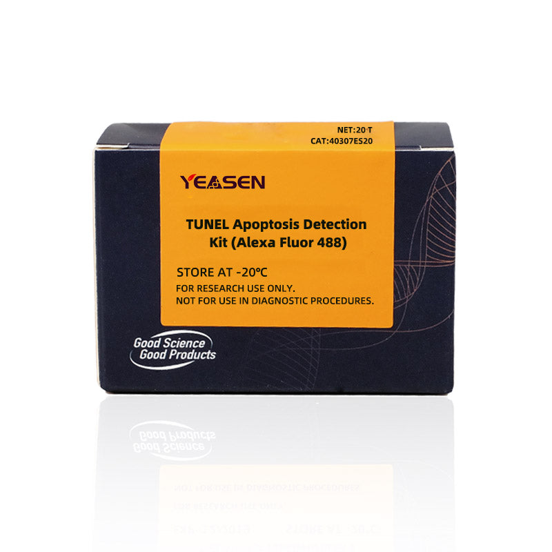
TUNEL (TdT mediated dUTP Nick End Labeling) Apoptosis detection kit (Alexa Fluor 488) can be used to detect nuclear DNA fragmentation in the late apoptotic process of tissue cells. The principle is that under the action of Terminal Deoxynucleotidyl Transferase (TdT), Alexa Fluor 488-12-DUTP is incorporated into the 3´ -hydroxyl (3´-OH) Terminal exposed during genomic DNA break. Thus, it can be detected by fluorescence microscopy or flow cytometry.
Alexa Fluor 488 dye is more stable and has a stronger signal, resulting in brighter markers and greater resistance to quench.
This kit has a wide range of applications and can be used to detect apoptosis in frozen or paraffin sections, as well as in cultured adherent or suspended cells.
Product Components
|
Component |
|
40307ES20(20 T) |
40307ES50(50 T) |
40307ES60(100 T) |
|
40307-A |
5×Equilibration Buffer |
750 μL |
1.25 mL×2 |
1.25 mL×3 |
|
40307-B |
Alexa Fluor 488-12-dUTP Labeling Mix |
100 μL |
250 μL |
250 μL×2 |
|
40307-C |
Recombinant TdT Enzyme |
20 μL |
50 μL |
50 μL×2 |
|
40307-D |
Proteinase K(2 mg/mL) |
40 μL |
100 μL |
100 μL×2 |
|
40307-E |
DNase I (1 U/μL) |
5 μL |
12.5 μL |
25 μL |
|
40307-F |
10 × DNase I Buffer with MgCl2 |
100 μL |
250 μL |
500 μL |
Shipping and Storage
The components are shipped with an ice pack and can be stored at -20°C for 1 year.
Cautions
1. It is necessary to prepare your own PBS for washing cells, an anti-fluorescence quenching solution for sealing tablets, and 4% paraformaldehyde for fixing.
2. For research use only!
Instructions
1. The sample preparation
1.1 Paraffin embedded tissue sections
1.1.1.The paraffin tissue sections were immersed in xylene for 5 min at room temperature and repeated once to remove paraffin thoroughly.
1.1.2.Soak the sections in 100% ethanol for 5 min at room temperature and repeat once.
1.1.3.At room temperature, the samples were soaked with gradient ethanol (90, 80, 70%) for 3 min each time.
1.1.4.Rinse the sections gently with PBS and carefully blot the excess liquid around the sample on the glass slides with filter paper. In this case, the paraffin pen or hydrophobic pen can be used to draw the outline of the sample distribution around the sample, which is convenient for downstream permeability processing and balance labeling operation. Do not let the sample dry during the experiment. Put the processed sample in the wet box to keep the sample wet.
1.1.5.2 mg/mL Proteinase K solution was diluted with PBS at a ratio of 1:100 to reach a final concentration of 20 μg/mL.
1.1.6.100 μL of Proteinase K solution at a concentration of 20 μg/mL was added to each sample to completely cover it and incubated at room temperature for 20 min.
[Notes]: Proteinase K helps tissues and cells become permeable to the staining reagents in subsequent steps. Too long incubation time will increase the risk of tissue sections falling off the carrier plate in subsequent washing steps, while too short incubation time may cause insufficient permeability treatment and affect labeling efficiency. To obtain better results, it may be necessary to optimize the incubation time of Proteinase K.
1.1.7.Rinse the sample 2-3 times with PBS solution, gently remove excess liquid, and carefully blot the liquid around the sample on the slide with filter paper. The processed sample is placed in a wet box to keep the sample moist.
1.2 Frozen section of tissue
1.2.1. Remove frozen sections and return to room temperature. The slides were immersed in 4% paraformaldehyde solution (dissolved in PBS) and fixed and incubated for 30 min at room temperature.
1.2.2. Gently remove excess liquid and use filter paper to carefully blot the excess liquid around the sample on the glass slide.
1.2.3. The slides were immersed in PBS solution, incubated at room temperature for 15 min, and washed with PBS again 2 times in total.
1.2.4. Gently remove excess liquid and carefully blot the glass slide with filter paper to remove excess liquid around the sample. In this case, the paraffin pen or hydrophobic pen can be used to draw the outline of the sample distribution around the sample, which is convenient for downstream permeability processing and balance labeling operation. During the experiment, do not let the sample dry, and put the processed sample in the wet box to keep the sample wet.
1.2.5. 2 mg/mL Proteinase K solution was diluted with PBS at a ratio of 1:100 to reach a final concentration of 20 μg/mL.
1.2.6. 100 μL of Proteinase K solution at a concentration of 20 μg/mL was added to each sample to completely cover it and incubated at room temperature for 10 min.
[Notes]: Proteinase K helps tissues and cells to be permeable to staining reagents in subsequent steps. Too long incubation time will increase the risk of tissue sections falling off the carrier plate in subsequent washing steps, while too short incubation time may cause insufficient permeability treatment and affect labeling efficiency. It may be necessary to optimize the incubation time of Proteinase K if better results were not obtained.
1.2.7. Rinse the sample 2-3 times in an open beaker containing PBS solution.
[Notes]: In order to avoid the loss of sample peeling in the cleaning step, it is recommended not to wash the bottle, but to soak the glass slides in PBS solution 2-3 times for cleaning.
1.2.8. Gently remove excess liquid and use filter paper to carefully blot the liquid around the sample on the slide. The processed sample is placed in a wet box to preserve the moisture of the sample.
1.3 Preparation of cell crawl sheet
Adherent cells were cultured on Lab-Tek of Chamber Slides. After apoptosis induction treatment, slides were washed twice with PBS.
1.4 Preparation of cell smears (Taking poly-lysine coated slides as an example)
1.4.1. The cells were resuspended in PBS at a concentration of about 2 × 10⁷ cells /mL, 50-100 μL of the cell suspension was aspirated onto poly-lysine coated slides, and the cell suspension was gently spread open with a clean slide.
1.4.2. The cells were fixed, and the slides were immersed in a staining tank containing 4% freshly prepared paraformaldehyde in PBS and placed at 4 ° C for 25 min.
1.4.3. The slides were washed, immersed in PBS, and left at room temperature for 5 min. Wash again with PBS.
1.4.4. Gently remove excess liquid and carefully blot the glass slide with filter paper to remove excess liquid around the sample. In this case, a paraffin pen or hydrophobic pen can be used to outline the distribution of the sample around the sample to facilitate downstream permeability processing and balance labeling operations. During the experiment, do not let the sample dry, and put the processed sample in the wet box to keep the sample wet.
1.4.5. 2 mg/mL Proteinase K solution was diluted with PBS at a ratio of 1:100 to reach a final concentration of 20 μg/mL.
1.4.6. 100 μL of Proteinase K solution at a concentration of 20 μg/mL was added to each sample to make it fully covered and incubated at room temperature for 5 min (it can also be immersed in 0.2% Triton X-100 solution prepared in PBS and incubated at room temperature for 5 min for permeability treatment).
[Notes]: Proteinase K helps tissues and cells to be permeable to staining reagents in subsequent steps. Too long incubation time will increase the risk of tissue sections falling off the carrier plate in subsequent washing steps, while too short incubation time may cause insufficient permeability treatment and affect labeling efficiency. It may be necessary to optimize the incubation time of Proteinase K if better results were not obtained.
1.4.7. Rinse the sample 2-3 times with PBS, gently remove excess liquid, and carefully blot the liquid around the sample on the slide with filter paper. The processed samples were placed in a wet box.
2. Steps for DNase treatment of positive controls
After sample permeation, cells were treated with DNase I to prepare positive control slides. This process usually causes most of the cells treated to show green fluorescence.
[Notes]: Dnase I treatment of immobilized cells causes breakage of chromosomal DNA, producing many 3 '-ends of DNA that can be labeled.
2.1.Dilute 10 × DNase I Buffer with deionized water at a ratio of 1:10 (200 µL 1× DNase I Buffer is required for each sample, that is, 20 µL 10 × DNase I Buffer and 180 µL deionized water are required for dilution). A drop of 100 µl was added to the permeable sample and incubated for 5min at room temperature. Add 1 μL of DNase I (1U/μL) to the remaining 100 μL of 1 × DNase I Buffer to achieve a final concentration of 10 U/mL.
2.2.The liquid was gently tapped off, then 100 μL of buffer containing 10 U/mL DNase I was added and incubated for 10 min at room temperature.
2.3.Tap the slide to remove excess liquid, and wash the slide thoroughly 3-4 times in a dye tank with deionized water.
[Notes]: A separate staining tank must be used for the positive control slides, otherwise residual DNase I on the positive control slides may introduce a high background on the experimental slides.
3. Labeling and Detection
3.1. Equilibration Buffer (5 × Equilibration Buffer) is diluted with deionized water in a ratio of 1:5 (100μL 1 × Equilibration Buffer is required for each sample).
3.2. Equilibration Buffer (100μL 1× Equilibration Buffer) was added to each sample to completely equilibrate the area and incubated for 10-30 min at RT. Alternatively, slide into a VAT with 1 x Equilibration Buffer to ensure that the slides are equilibrated. Thaw Alexa Fluor 488-12-DUTP Labeling Mix on ice while balancing cells, and prepare enough TdT incubation buffer for all experiments and optional positive control reactions according to Table 1. For a standard reaction with an area of fewer than 5 cm2, the volume is 50 μl, and 50 μl is multiplied by the number of experimental and positive control reactions to determine the total volume of TdT incubation buffer required. For samples with larger surface areas, the reagent volume can be increased proportionally.
Table 1 TdT incubation buffers prepared for experiments and optional positive control reactions
|
component |
volume(μL/50 μL system) |
|
ddH2O |
34 |
|
5×Equilibration Buffer |
10 |
|
Alexa Fluor 488-12-dUTP Labeling Mix |
5 |
|
Recombinant TdT Enzyme |
1 |
[Negative control system] : A control incubation buffer without TdT enzyme was prepared and the TdT enzyme was replaced with ddH2O.
3.3. Most of the 100 μl 1× Equilibration Buffer was washed off with absorbent paper around the equilibrated area and then 50 μl TdT incubation Buffer was added to a 5 cm2 area of cells. Don't let the cells dry out. After this, the slide should be shielded from light.
3.4. Place a plastic coverslip over the cells to ensure an even distribution of the reagents, and place a paper towel dampened with water at the bottom of the wet box. The slides were placed in a wet box and incubated at 37 ℃ for 60 min. Wrap the wet box in aluminum foil to protect it from light.
[Notes]: The plastic cover glass can be cut in half before use. Fold the edge of the cover glass for easy removal and manipulation.
3.5. The plastic coverslips were removed, and the sections were incubated in PBS solution at room temperature for 5 min. Then the sections were washed twice with fresh PBS.
3.6. Gently wipe the PBS solution around and on the back of the sample with filter paper.
[Notes]: In order to reduce the background, after washing the slides with PBS once, they can be washed with PBS containing 0.1% Triton X-100 and 5 mg/mL BSA 3 times, 5 min each time, so that the free unreacted markers can be clear and clean.
3.7. Samples were stained in a staining tank, and slides were immersed in a staining tank containing PI solution (1 μg/mL, freshly prepared and diluted with PBS) in the dark and left at room temperature for 5 min. (Optional): Samples were stained in a staining tank, and slides were immersed in a staining tank containing DAPI solution (2 μg/mL, freshly prepared and diluted with PBS) in the dark, and left at room temperature for 5 min.
3.8. Wash the sample, immerse the slide in deionized water and leave it at room temperature for 5 minutes. Repeat twice for a total of three washes.
3.9. The excess water on the slide was knocked dry and 100 μl PBS was added to the sample area to keep the sample moist.
3.10. The samples were immediately analyzed under a fluorescence microscope, and the green fluorescence was observed at 520±20 nm with a standard fluorescence filter device. The red fluorescence of PI was observed at > 620 nm, or the blue DAPI was observed at 460 nm. If necessary, the slides can be stored overnight at 4 ° C in the dark.
[Notes]: PI/DAPI could stain both apoptotic and non-apoptotic cells red/blue, and only in apoptotic nuclei was Alexa Fluor 488-12-DUTP incorporated and localized green fluorescence.
4. The suspension cells were detected by flow cytometry
4.1. 3~5 × 10⁶ cells were washed twice with PBS by centrifugation (300 × g) at 4 ° C, centrifuged at 300 g at 4 ° C for 10 min, and then resuspended in 0.5 mL PBS.
4.2. The cells were fixed, and 5 mL of 1% paraformaldehyde solution prepared in PBS was added and placed on ice for 20 min.
4.3. Cells were centrifuged at 300 × g at 4 ° C for 10 min, and the supernatant was removed and resuspended in 5 mL PBS. The wash was repeated once and the cells were resuspended with 0.5 mL PBS.
4.4. The cells were permeated, and 5 mL of ice-pre-cooled 70% ethanol was added and incubated at -20 ° C for 4 hours. The cells could be stored in 70% ethanol at -20℃ for a week, or the cells could be permeable with 0.2% Triton X-100 solution prepared in PBS and stored at room temperature for 5 min.
4.5. Cells were centrifuged at 300 × g for 10 min and resuspended in 5 mL PBS. Centrifugation was repeated and resuspended in 1 mL PBS.
4.6. Transfer 2×10⁶ cells to a 1.5-ml microcentrifuge tube.
4.7. Equilibration was centrifuged at 300×g for 10 min, and the supernatant was removed and resuspended with 80 μL 1×Equilibration Buffer. Incubate at room temperature for 5 min.
4.8. While balancing cells, Alexa Fluor 488-12-DUTP Labeling Mix was melted on ice, and enough TdT incubation buffer for all reactions was prepared according to Table 1. For a standard reaction of 2×10⁶ cells, the volume is 50 μL, and 50 μL times the number of reactions is the total volume of TdT incubation buffer required.
4.9. The cells were centrifuged at 300×g for 10 min, the supernatant was removed and the precipitate was resuspended in 50μL TdT incubation buffer and incubated at 37℃ for 60 min, shielded from light. Cells were gently resuspended every 15 min with a micropipette.
4.10. The reaction was terminated by adding 1 mL of 20 mM EDTA and gently mixing with a micropipette.
4.11. After centrifugation at 300g for 10 min, the supernatant was discarded and the precipitate was resuspended in 1 mL 0.1% Triton X-100 solution prepared in PBS, containing 5 mg/mL BSA. The solution was repeated once and washed twice in total.
4.12. The supernatant was discarded by centrifugation at 300 g for 10 min, and the cells were resuspended in 0.5 mL of 5μg/mLPI solution newly prepared with PBS, which contained 250 μg of DNase-free RNase A.
4.13. Cells were incubated in the dark for 30 min at room temperature.
4.14. The cells were analyzed by flow cytometry, and the green fluorescence was observed at 520±20 nm. The red fluorescence of PI was observed at > 620 nm. PI could stain both apoptotic and non-apoptotic cells red, and only in apoptotic nuclei was Alexa Fluor 488-12-DUTP incorporated and localized green fluorescence.
.
Payment & Security
Your payment information is processed securely. We do not store credit card details nor have access to your credit card information.
Inquiry
You may also like
FAQ
The product is for research purposes only and is not intended for therapeutic or diagnostic use in humans or animals. Products and content are protected by patents, trademarks, and copyrights owned by Yeasen Biotechnology. Trademark symbols indicate the country of origin, not necessarily registration in all regions.
Certain applications may require additional third-party intellectual property rights.
Yeasen is dedicated to ethical science, believing our research should address critical questions while ensuring safety and ethical standards.

